About the book
This bestselling textbook continues to provide a broad introduction to the key topics in the welfare of animals both large and small, farm and companion, wild and zoo. It retains all the popular features of the previous editions with coverage of key issues such as ethics, animal pain and injury, health and disease, social conditions, and welfare dilemmas and problems. Importantly, it also offers practical advice for welfare assessment, with a full section dedicated to the implementation of solutions. The third edition:
- Contains many more examples of welfare issues in different countries, particularly the implications for smallholders as well as larger scale agriculture
- Covers fish welfare as well as welfare of amphibians, reptiles and invertebrates
- Includes concepts of positive emotion and other positive aspects of welfare
- Focuses on animal welfare and sustainability
- Includes an integrated eBook with additional material and videos
Access the additional online content and videos below.


‘…ample research has now shown that the traditional battery cage for laying hens frustrates the hen’s motivation to perform dust-bathing behaviour (Video 2.1), thus breaching the ‘freedom’ to perform normal behaviour.’
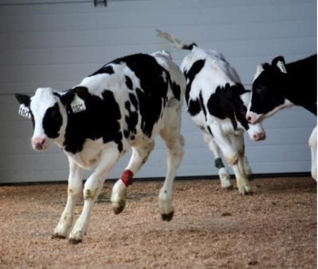
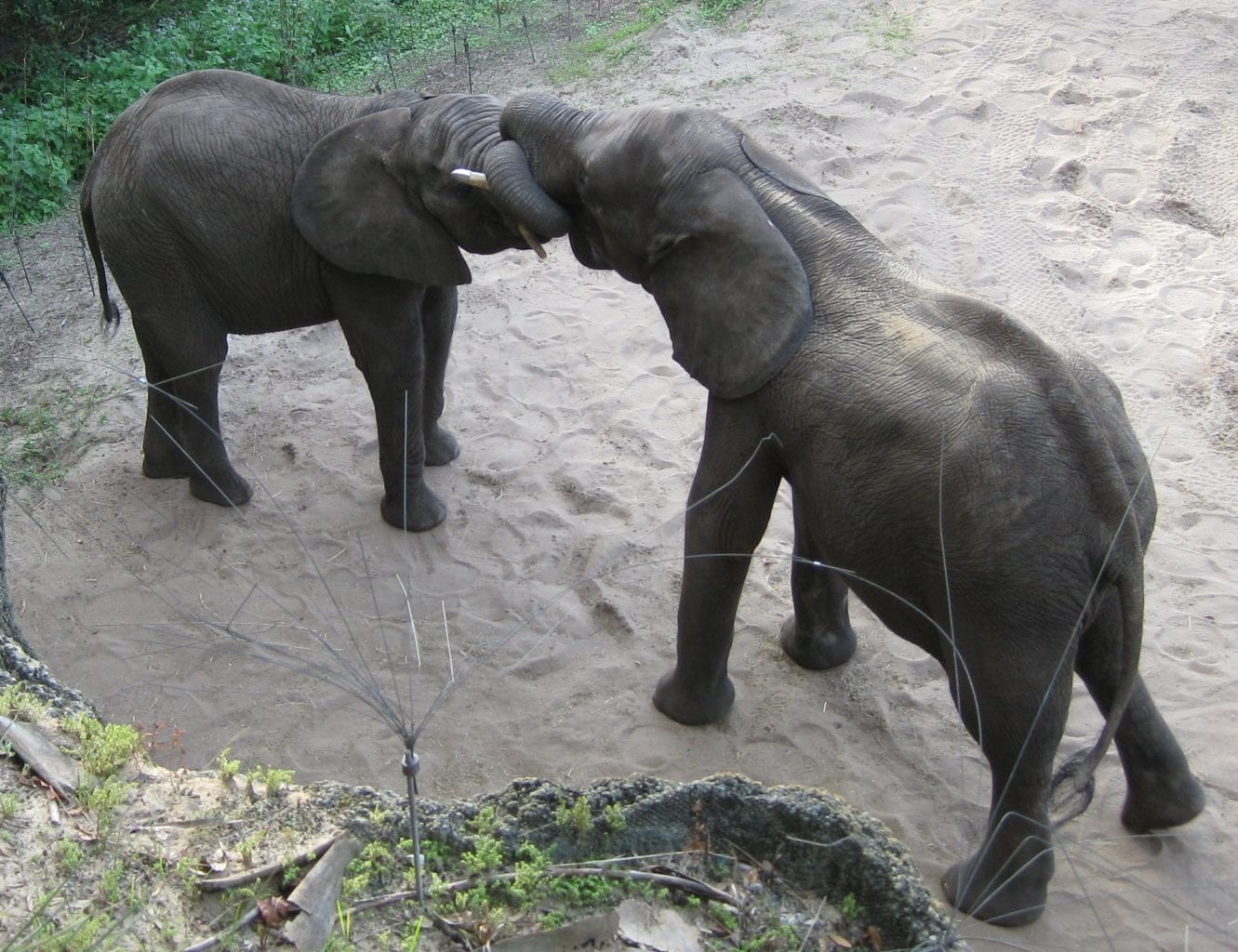
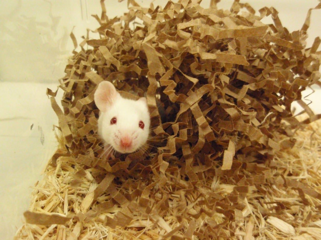
‘Perhaps one of the biggest shifts is towards finding signs that may indicate that the welfare of an animal is good, rather than signs that its welfare is bad, and in investigating positive emotions (Boissy et al., 2007a). Among the best-established indicators is the presence of play behaviour (Held and Špinka, 2011), although mainly applicable in young animals (Fig. 2.9E, Videos 2.2 and 2.3).’ Courtesy of Yezica Norling.
‘Perhaps one of the biggest shifts is towards finding signs that may indicate that the welfare of an animal is good, rather than signs that its welfare is bad, and in investigating positive emotions (Boissy et al., 2007a). Among the best-established indicators is the presence of play behaviour (Held and Špinka, 2011), although mainly applicable in young animals (Fig. 2.9E, Videos 2.2 and 2.3).’ Courtesy of Yezica Norling.
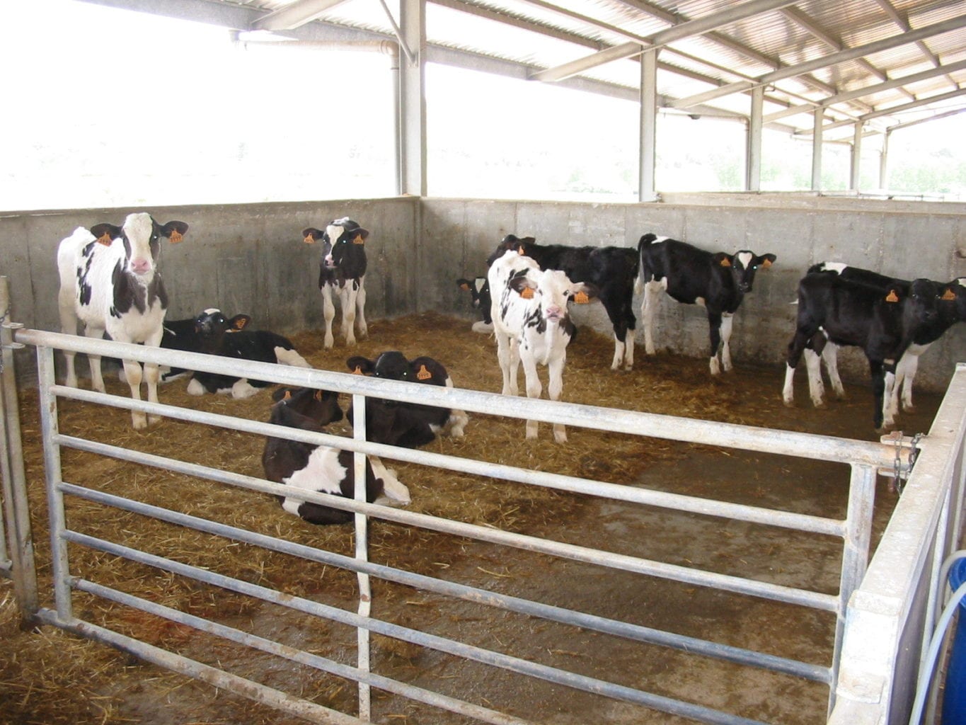
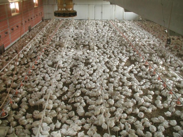
‘The Brambell Committee drew particular attention to the problems of behavioural restriction and frustration that resulted from intensive, indoor housing systems for farm animals (Video 2.4).’
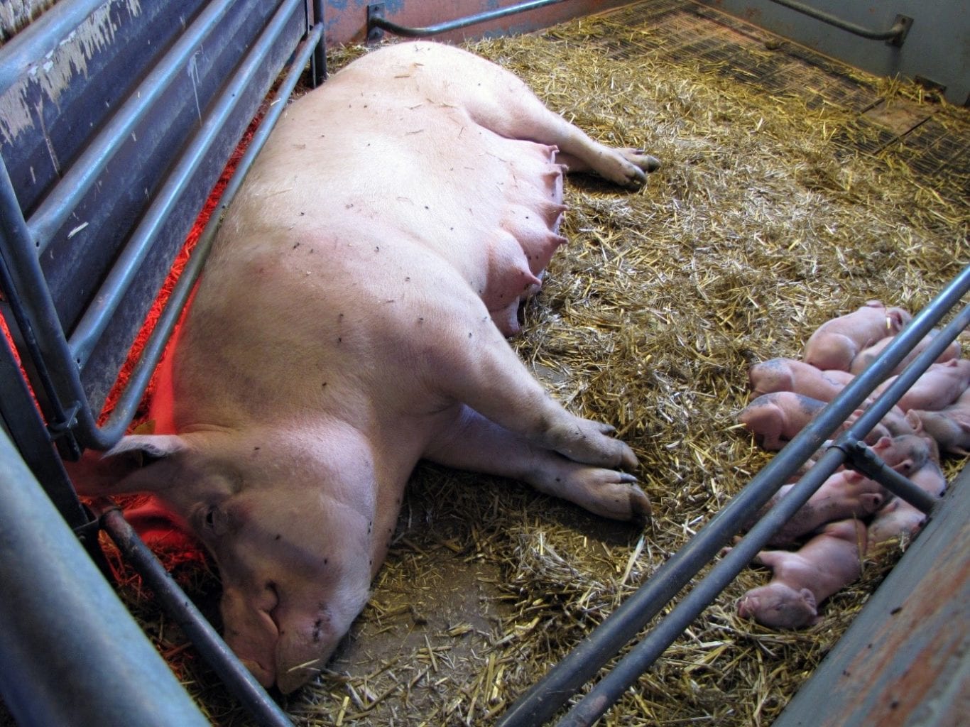
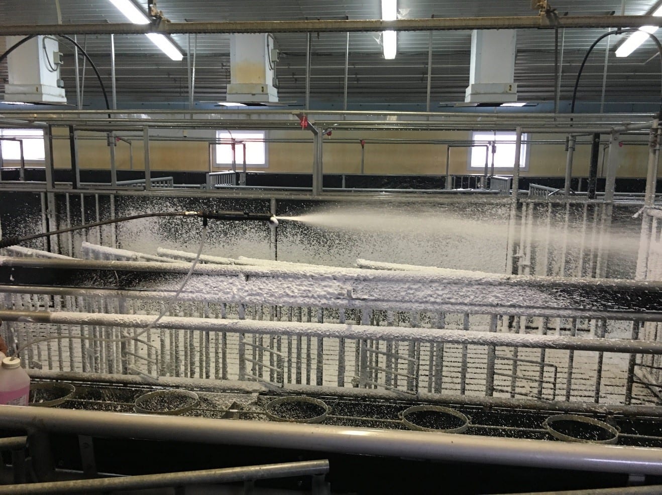
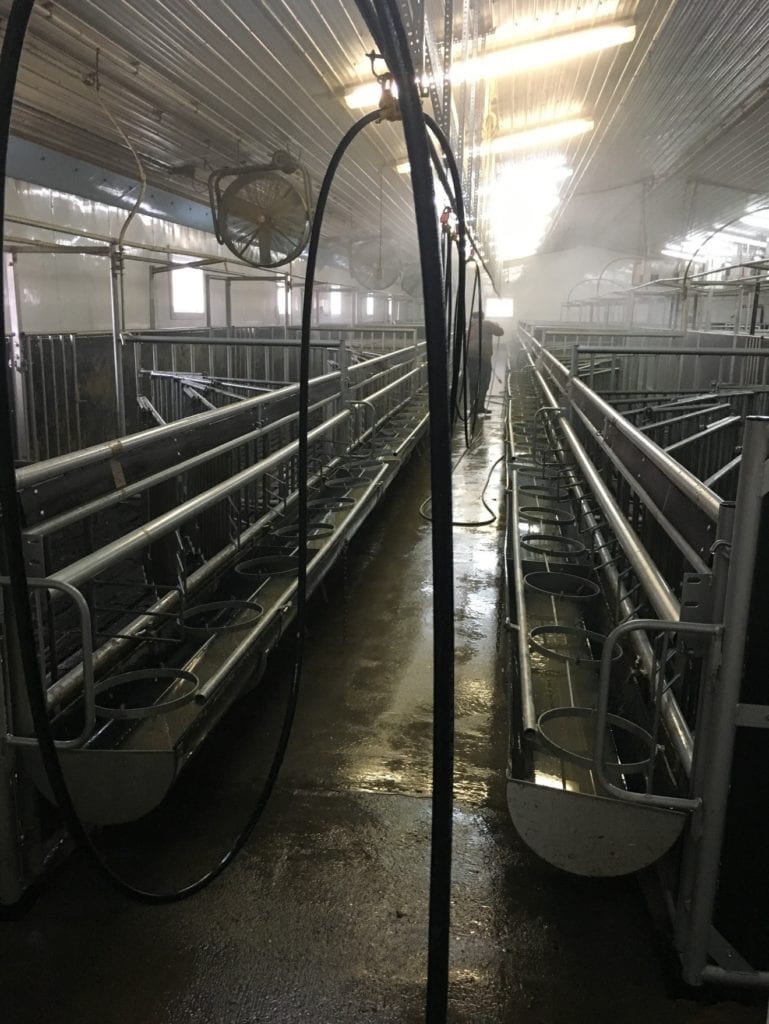
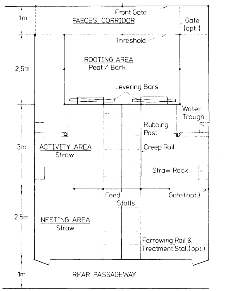
Welfare issues of pregnant and lactating sows.
Transcript
Welfare issues of farrowing sows and piglets.
Transcript
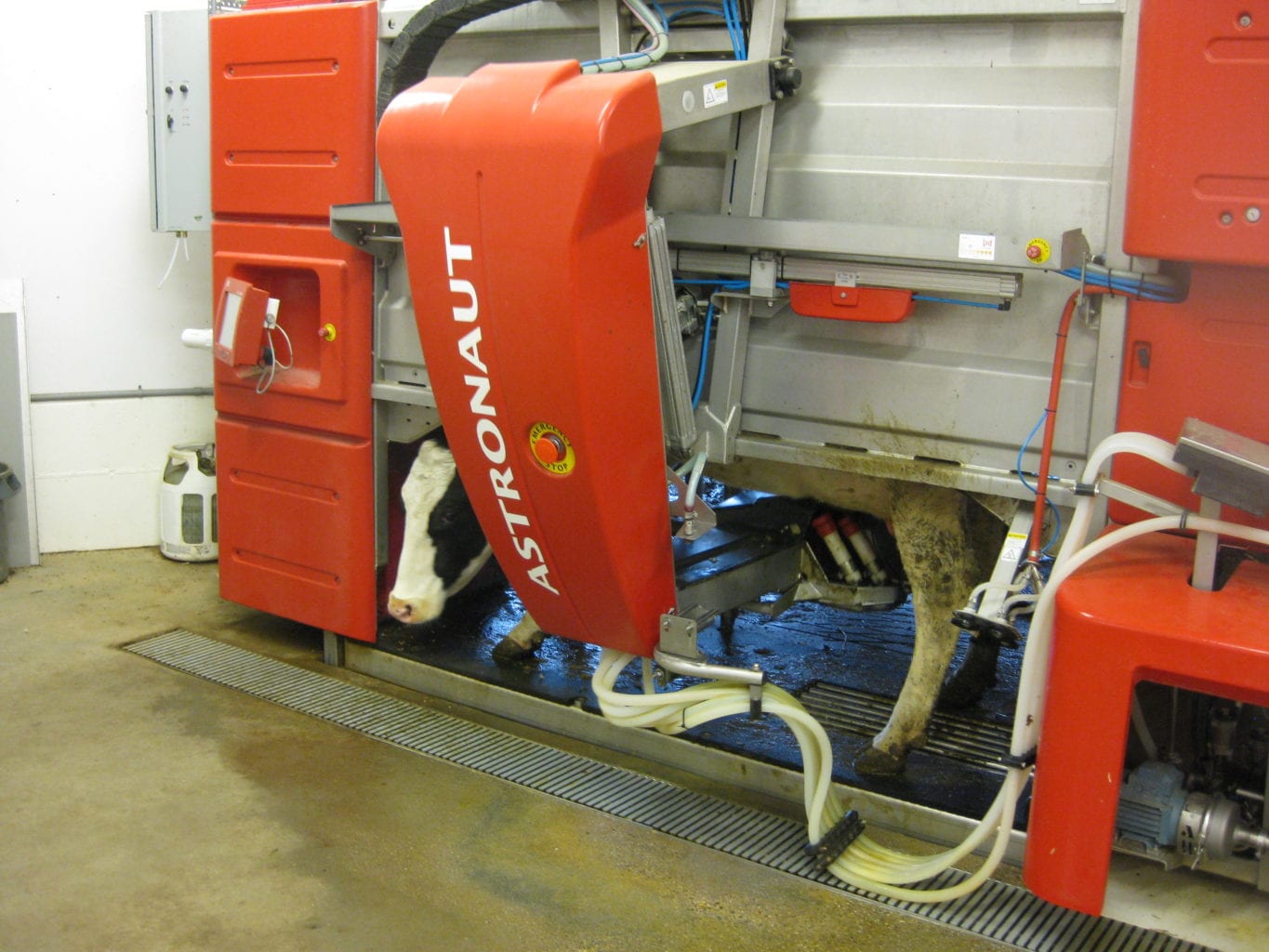
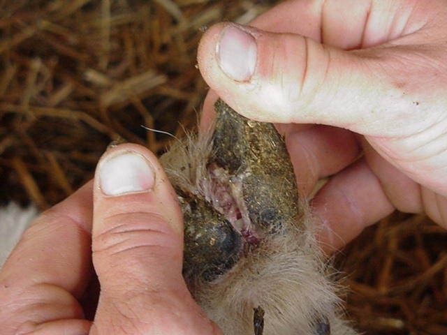
‘A considerable amount is known about environmental design and management in relation to the reduction or avoidance of specific physical welfare problems of injury, disease and discomfort (Video 13.1). This is particularly true in agriculture, where many such problems reduce profit.’ Video courtesy of Fritha Langford, University of Edinburgh.
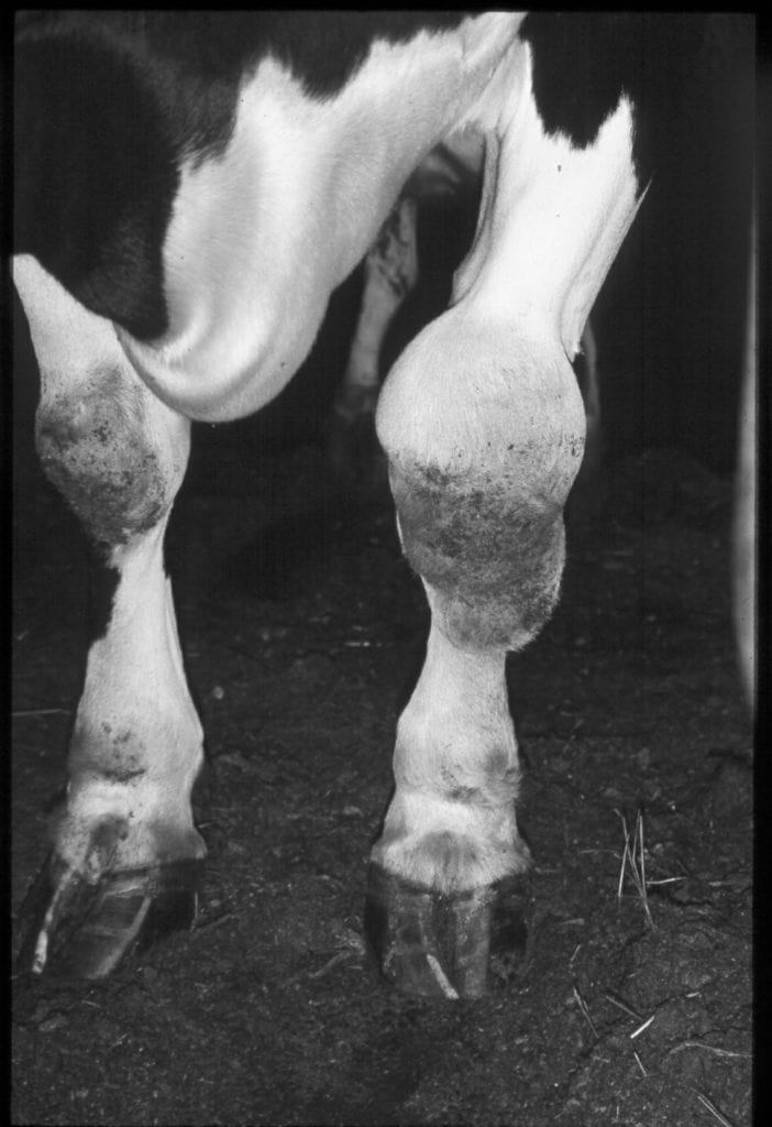
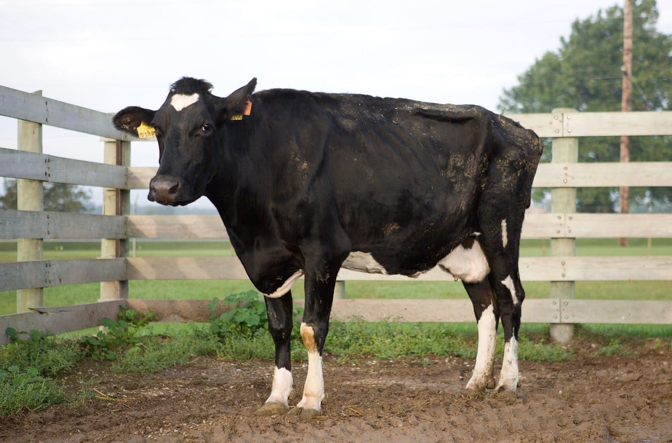
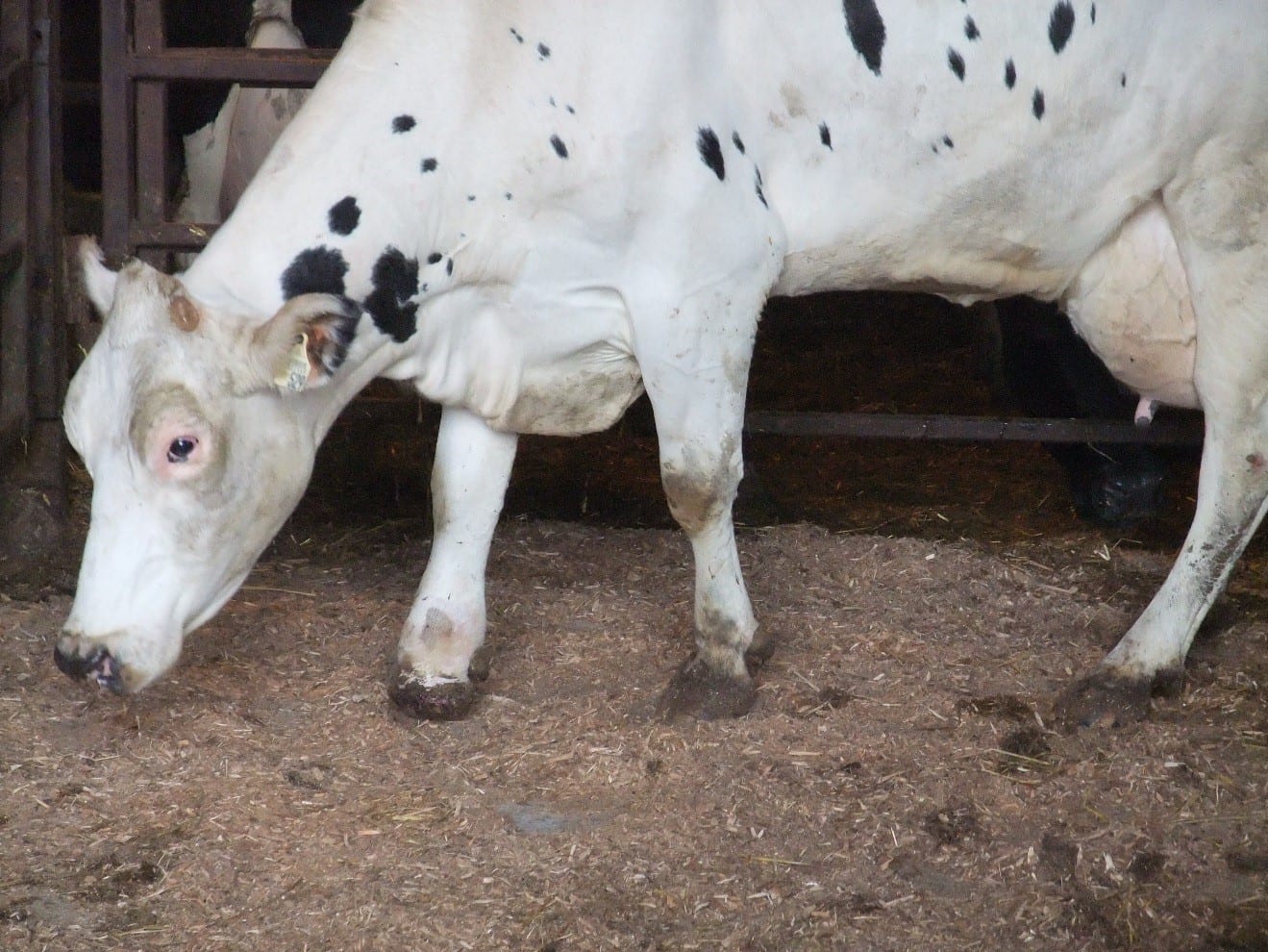
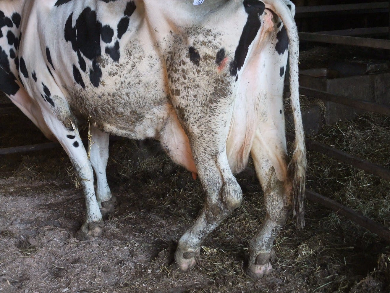
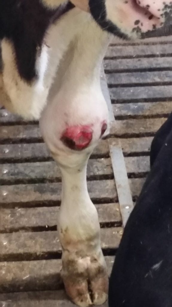
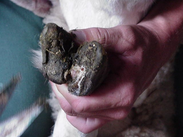
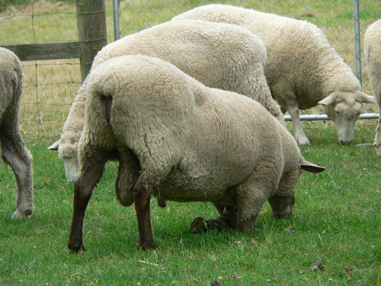
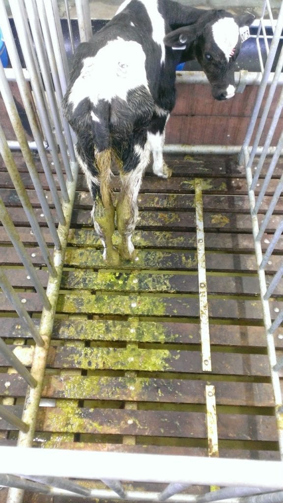
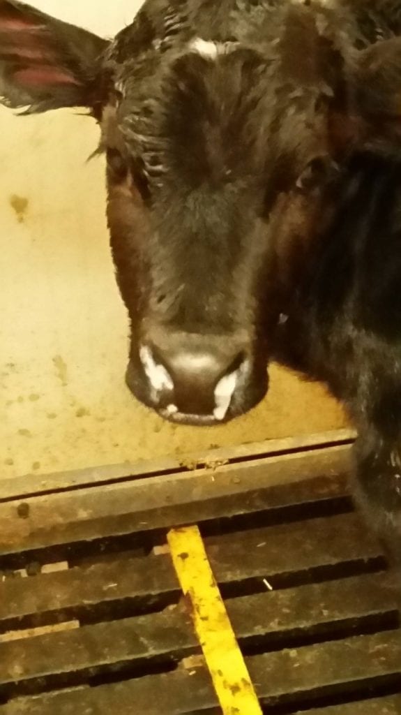
'Cows in cubicle housing: bedding is provided in cubicles, but one cow stands ruminating on wet, faeces-covered concrete and others stand with forefeet on bedding, hind feet on concrete. Video courtesy of Fritha Langford, University of Edinburgh. ‘…in the prevention of injury, the softness of the floor is important for both pigs (Lewis et al., 2005) and cattle (Frankena et al., 2009). However, the provision of appropriate floors to reduce lameness is not straightforward (Video 13.2).’
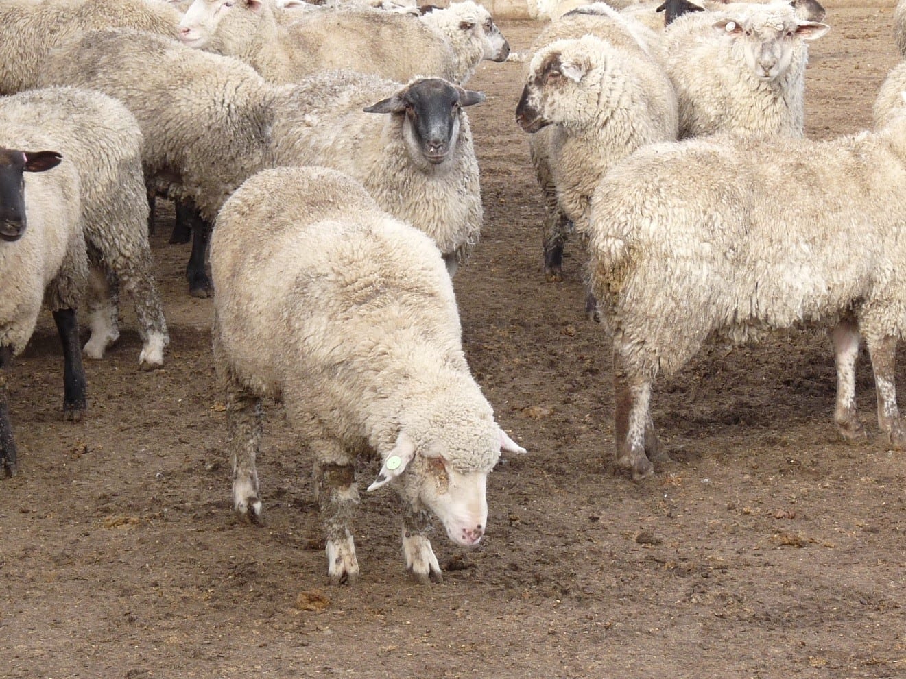
‘There are two major reservations to make about measures to reduce injury, disease and discomfort. Sometimes, measures known to be effective are not used, for various reasons including economics (Video 13.3), as emphasized in Chapter 8 (this volume) on health and disease. In contrast, some measures may be adopted irrespective of their deleterious effects on other aspects of welfare.’ Video courtesy of Fritha Langford, University of Edinburgh.
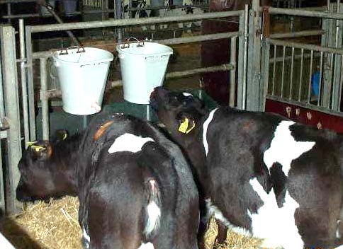
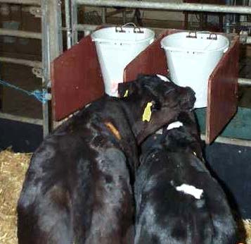
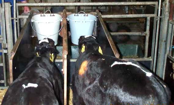
‘Sometimes, modification of the environment may not only lead to a reduction in undesirable food-related behaviour but also may affect production-related measures positively. This is the case when increased feeding space leads to a decrease in aggression and more time spent feeding, especially for low-ranking individuals, as, for example, in goats (Jørgensen et al., 2007) and cows (Huzzey et al., 2006) (Video 13.4).’ Video courtesy of Fritha Langford, University of Edinburgh.
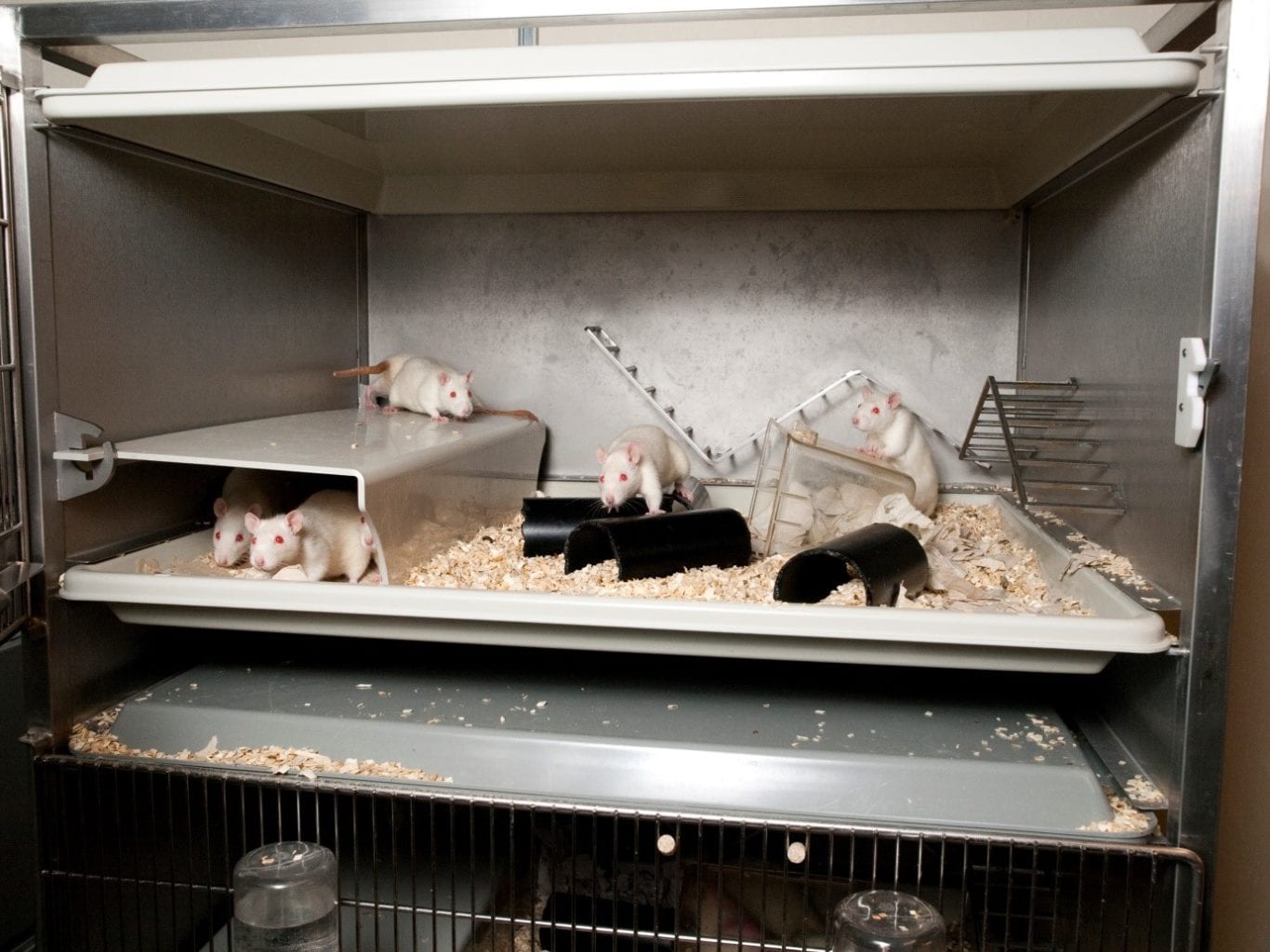
‘…trials (and others by other research groups) formed the basis for furnished or enriched cages (Video 13.5), which since 2012 have been the only type of cages permitted for use for laying hens within the EU.’ Presented by Vicky Sandilands and used with her permission.
Transcript
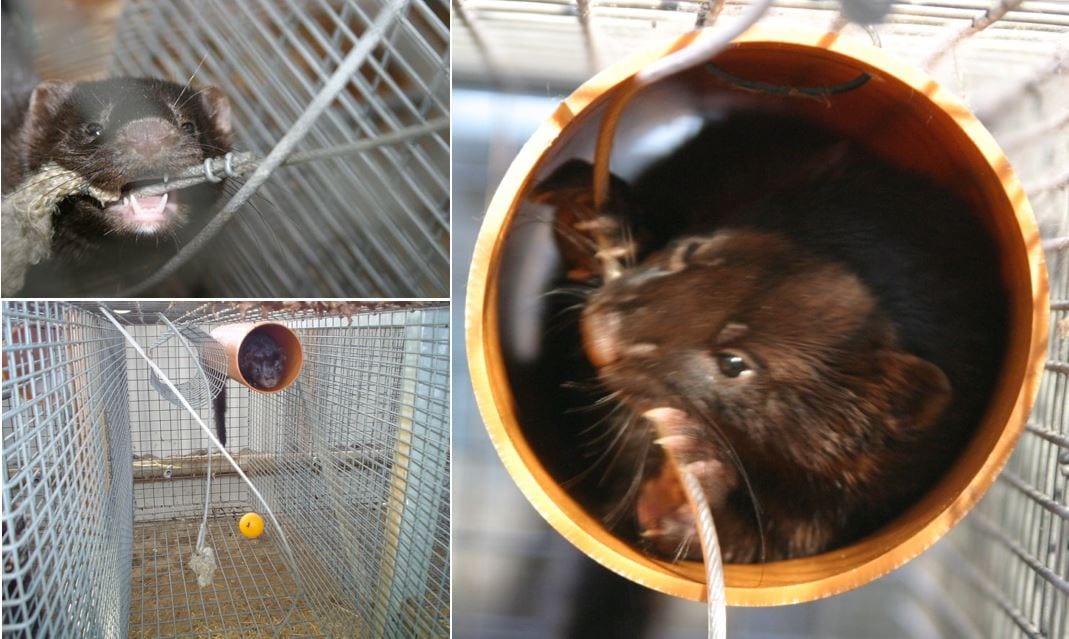
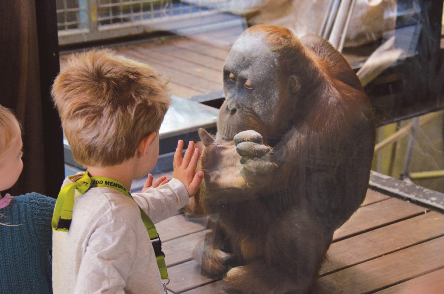
Not all zoos provide such enrichments. Their reasons may include: (a) concern that such objects and behaviour appear ‘unnatural’; (b) the cost of providing space and pools and keeping them clean; and (c) pressure on keepers’ time. However, such factors are increasingly outweighed by expectations from visitors and others that the welfare of animals in the zoo should be maximized. (Courtesy of Fritha Langford, University of Edinburgh.)
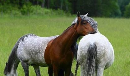
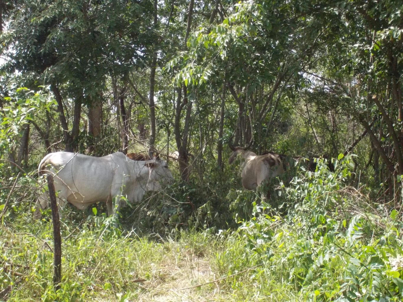
‘Améndola et al. (2016) found that the shade and microclimate (a reduction of 3–4°C in comparison to monoculture systems has been reported), as well as the feeding resources associated with these systems, resulted in less dispersed groups of cows, more affiliative interactions between cows and more stable hierarchies in comparison to monoculture systems (Video 14.1).’
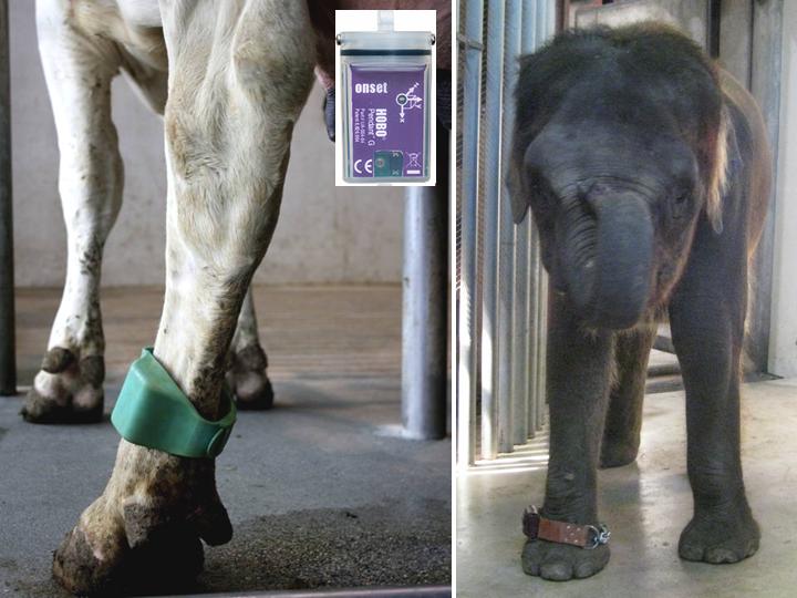
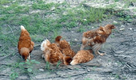
Lines diverging very significantly in their motivation for spontaneous locomotor behaviour in the home pen have been developed by genetic selection using the data obtained when signals from the individual electronic transponder attached to the leg of each chicken are received by the antennae, the white rectangular boxes in the litter (Kjaer, 2017). (Video recorded at FLI, Celle, by Joergen Kjaer and Oliver Sanders.)
More about CABI books

Open resources
A number of CABI’s books are enhanced by open resources. These materials are available for students and…